


Soft Tissue Grafts
CASE 22
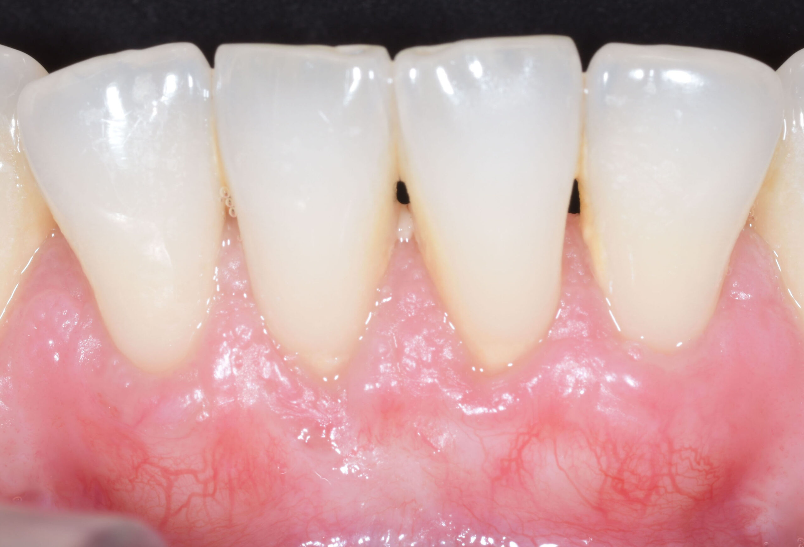
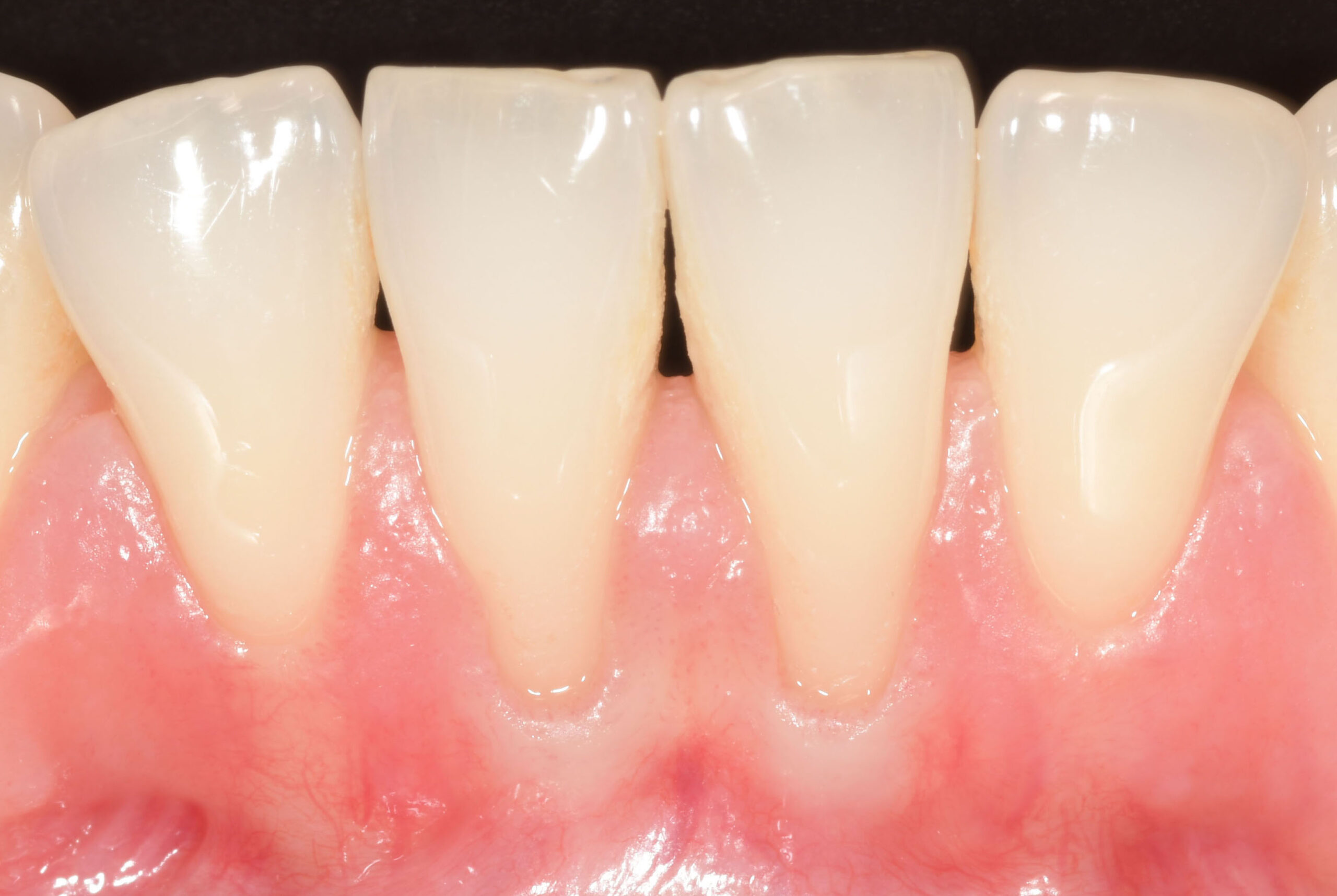
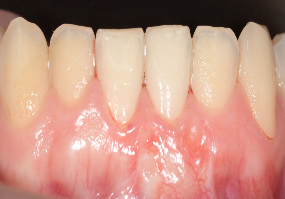
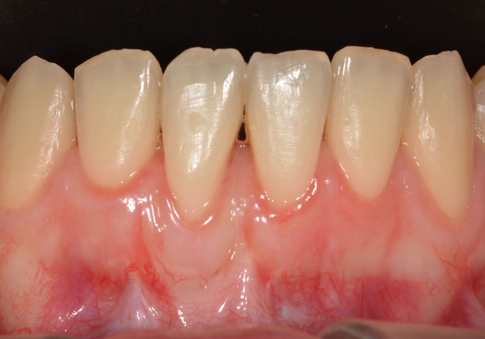
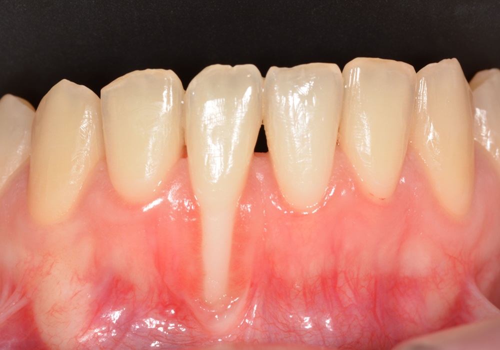
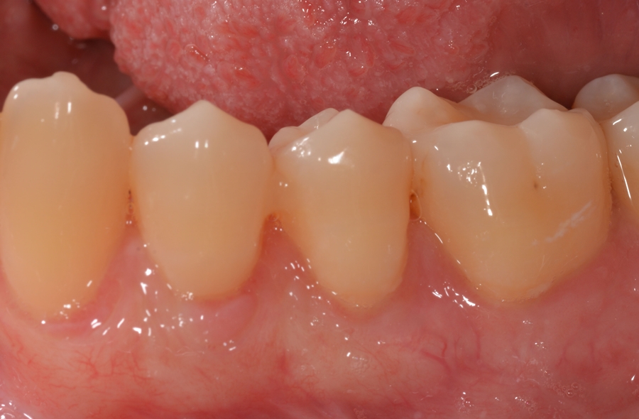
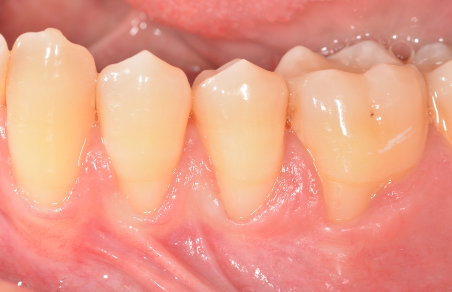
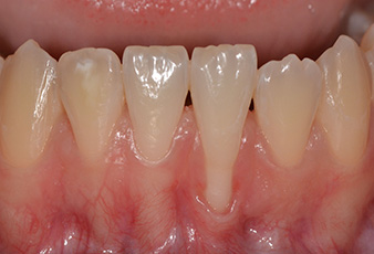
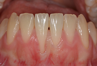
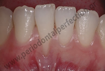
Diagnosis: Root recession in the 2 central lower incisors as well as inadequate keratinized gingiva that made painful the practicing of oral hygiene.
Treatment: The technique chosen was a vertically positioned flap combined with an autogenous deepithelialized free gingival graft that gave an impressive root coverage result.
CASE 21
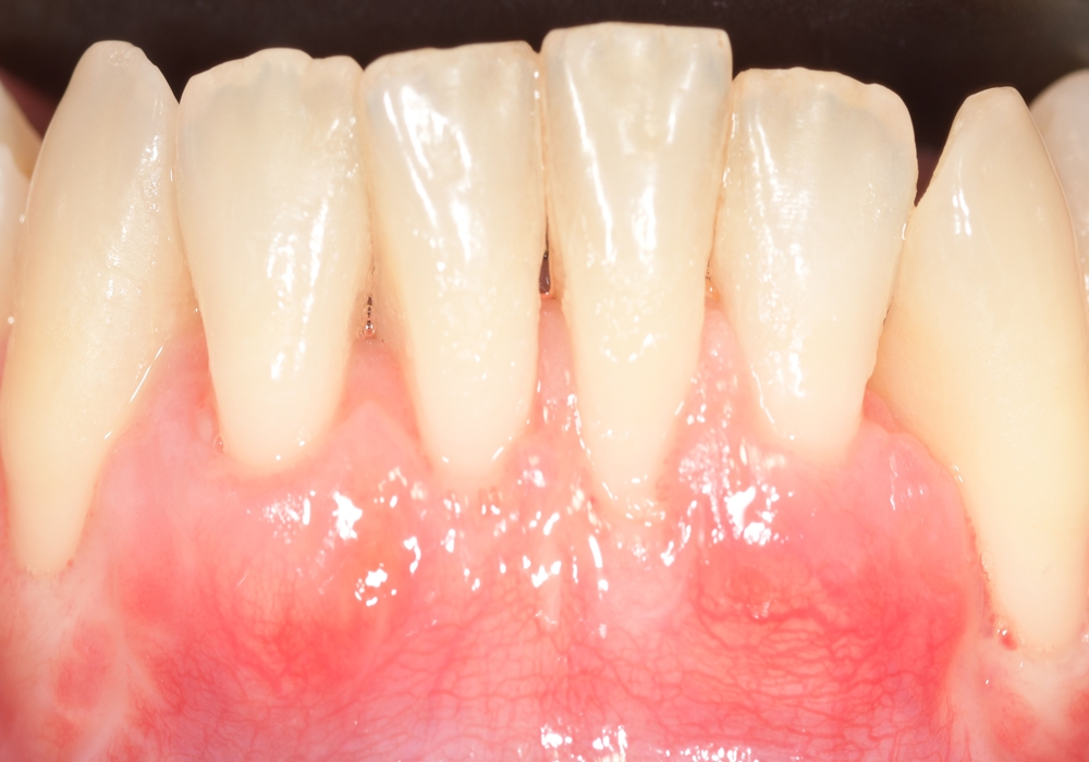
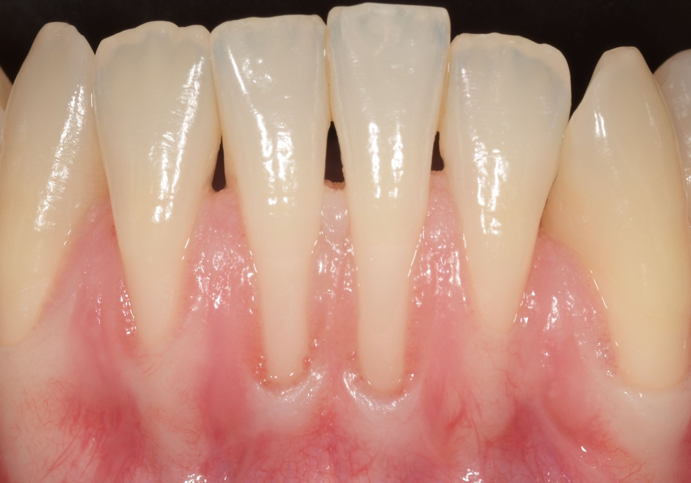
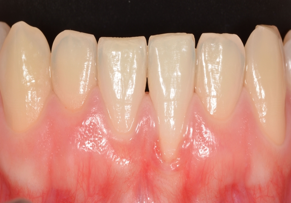
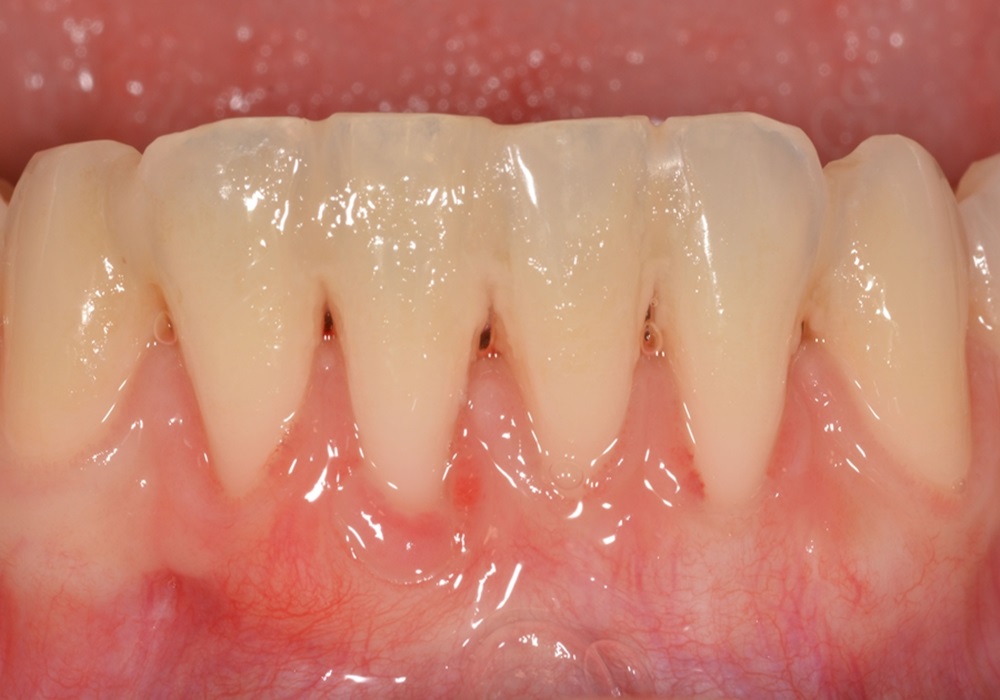
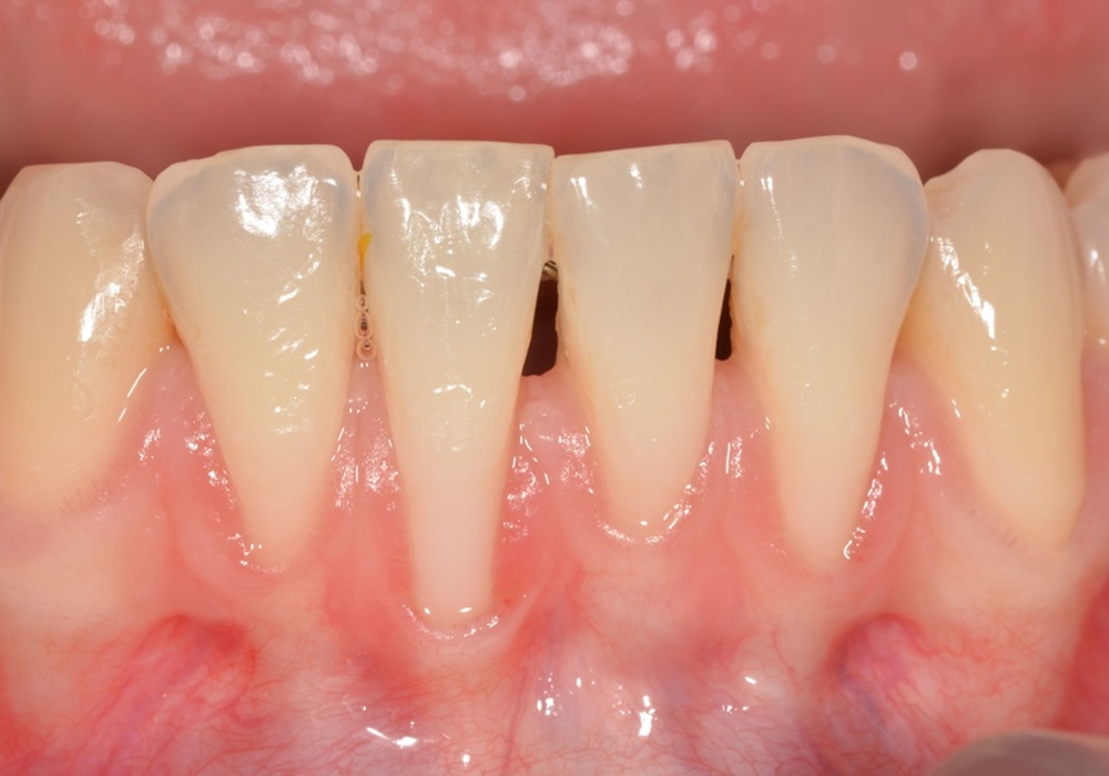
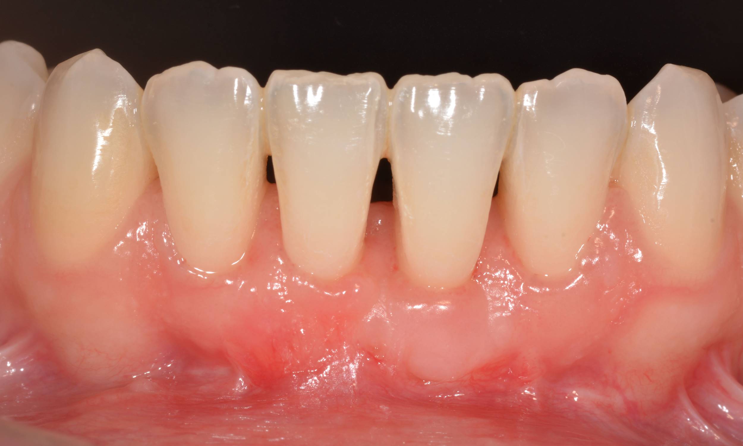
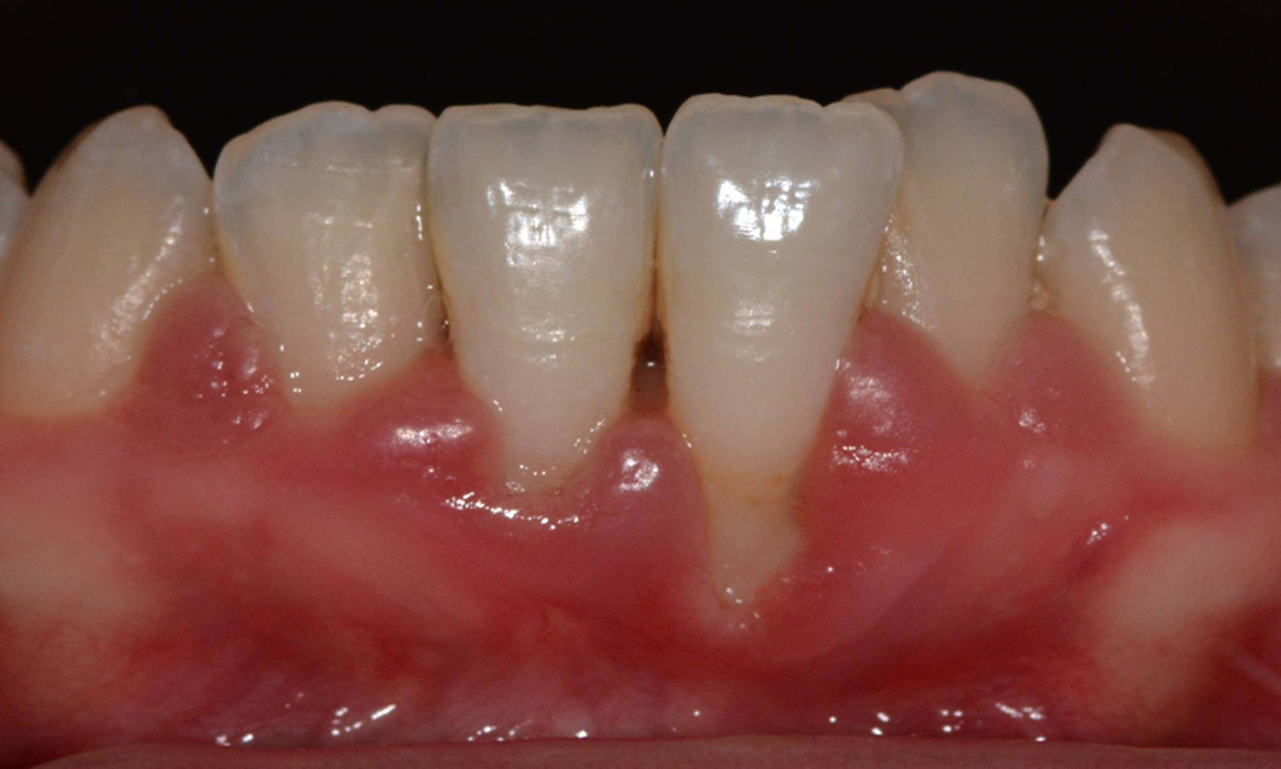
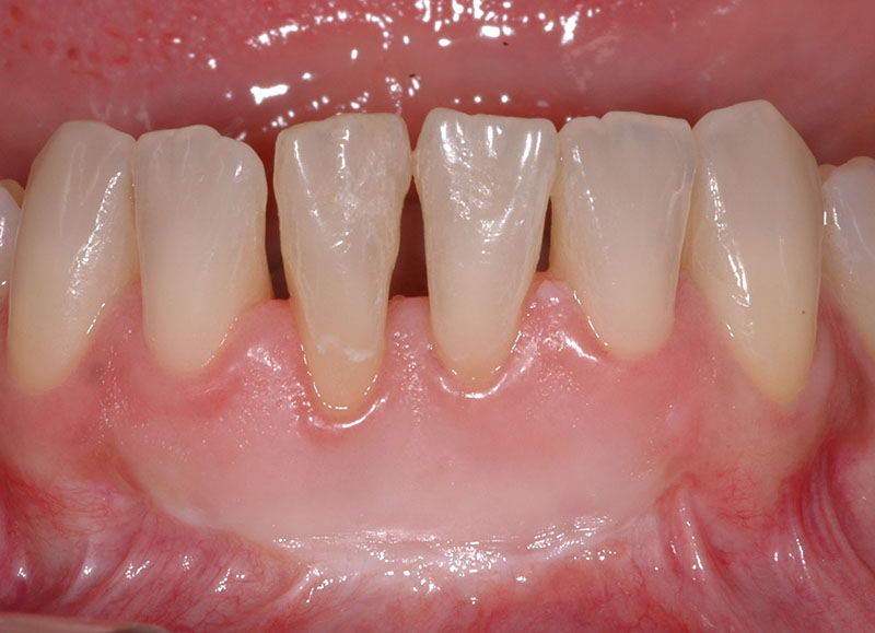
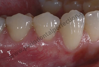
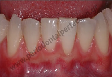
Diagnosis: Root recession in the 4 lower incisors classified as a Miller class III, which implies the inability of achieving full coverage after soft tissue grafting.
Treatment: The technique chosen was a vertically positioned flap combined with an autogenous deepithelialized free gingival graft that gave almost 100% root coverage as seen in the photograph.
CASE 20
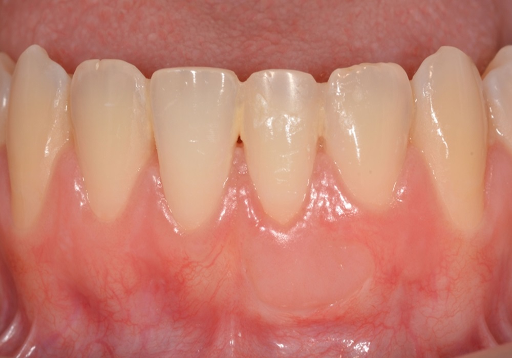
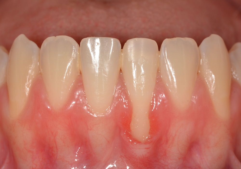
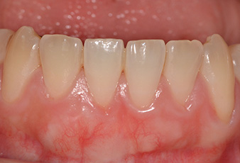
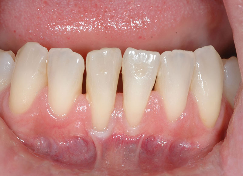
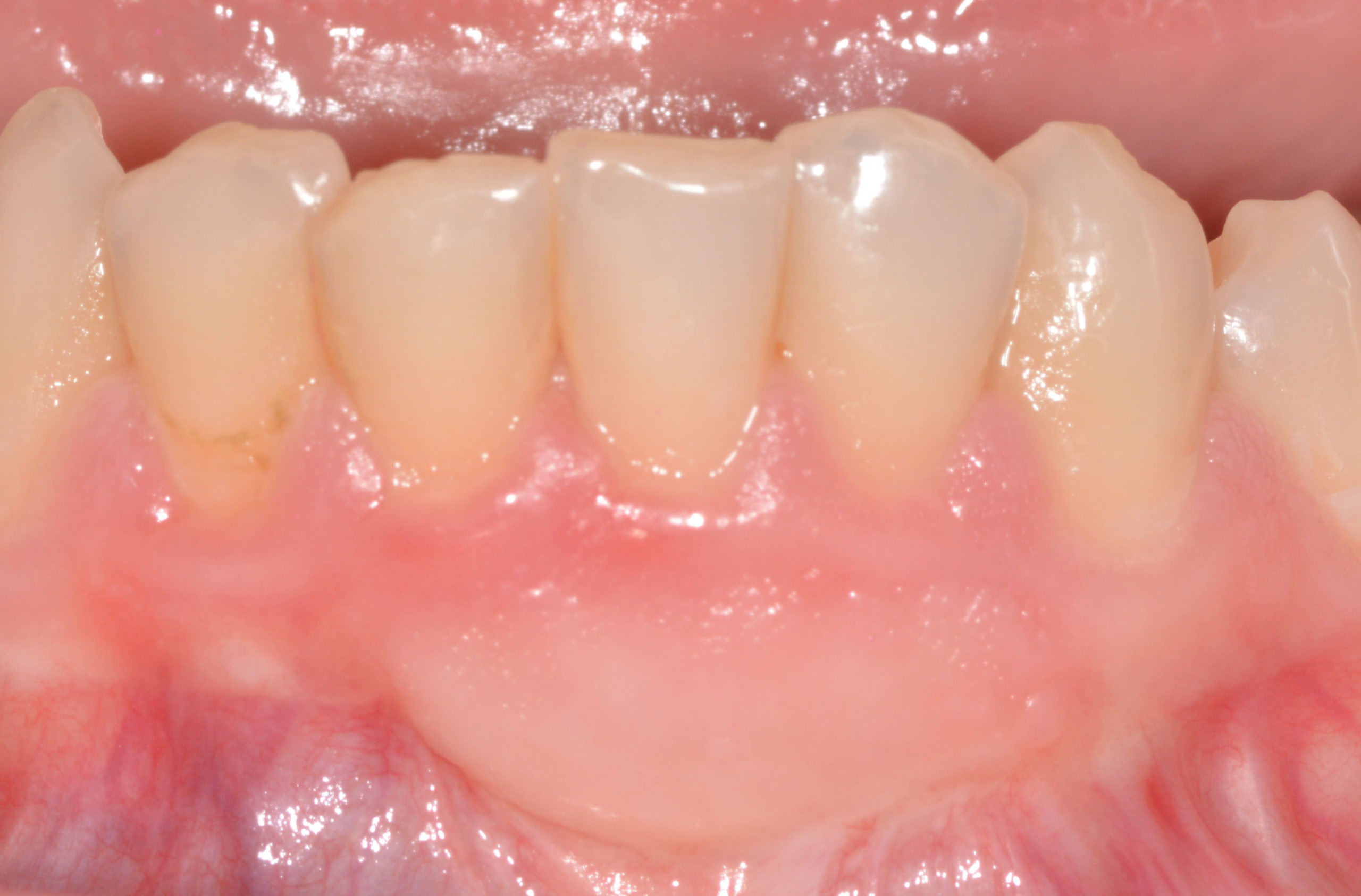
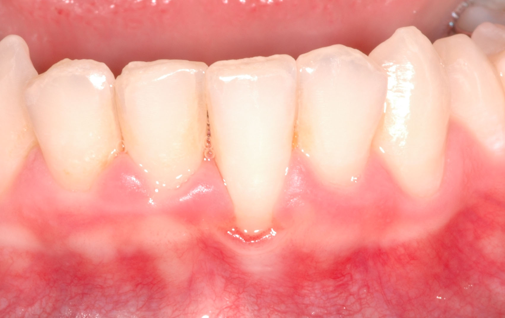
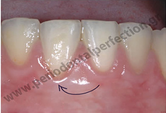
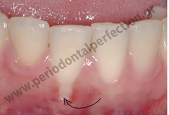
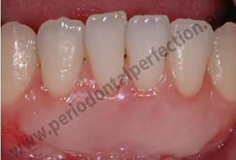
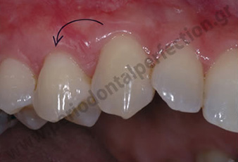
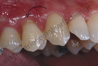
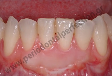
Diagnosis:: Υφίζηση στον αριστερό κεντρικό τομέα της κάτω γνάθου κατηγορίας Ι κατά Miller κάτι που συνεπάγεται τη δυνατότητα επίτευξης πλήρους κάλυψης μετά από ουλικό μόσχευμα.
Treatment:The technique chosen was a vertically positioned flap combined with an autogenous deepithelialized free gingival graft that gave 100% root coverage as seen in the photograph 2 months later.
ΠΕΡΙΣΤΑΤΙΚΟ 19
Diagnosis::
Treatment::
ΠΕΡΙΣΤΑΤΙΚΟ 18
Diagnosis:
Treatment:
ΠΕΡΙΣΤΑΤΙΚΟ 17
Diagnosis: Root recessions in all four lower incisors with the most pronounced being the recession in the central incisor of the right side of the patient. At the same time there is bone loss interdentally and that classifies the case to a Miller class III, which implies the inability to achieve full coverage after soft tissue grafting.
Treatment: The technique chosen was an autogenous deepithelialized free gingival graft placed in the recipient cite through a tunnel. The root coverage although not complete, was very impressive a few months later.
CASE 16
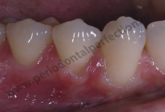
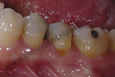
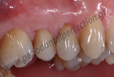
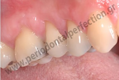
Diagnosis:Root recession from canine to first molar in the lower left sextant.
Treatment:The treatment provided to this patient was an autogenous soft tissue graft through a tunnel, meaning no incisions in the papillae. The percentage of root coverage achieved was 100% as shown in the picture taken 6 months later.
CASE 15
Diagnosis: Diagnosis: Root recession in the central incisors of the lower jaw and buccal inclination of those teeth. In this case, the presence of microbial dental plaque and heavy deposits of calculus (tartar) are obvious as well as the gingival inflammation.
Treatment: Α) The patient initially underwent conservative periodontal treatment that included oral hygiene instructions and control of inflammation by removing microbial plaque. B) Then orthodontic treatment was proposed to adjust the incisors in a more favorable position inside the mandible so as to increase the percentages of the expected root coverage. C) Six months after the completion of the orthodontic treatment, we proceeded to an autogenous soft tissue graft in order to cover the recessions and the technique used was the vertically positioned flap. In this case amelogenin was also used to improve the quality of connective tissue attachment.
CASE 14
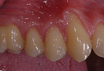
Diagnosis: Root recessions in the lateral incisor, canine and first premolar upper left.
Treatment: The technique utilized is the one described by Zucchelli for the upper jaw and the root coverage reached 100%. The photo on the right was taken 6 months later.
CASE 13
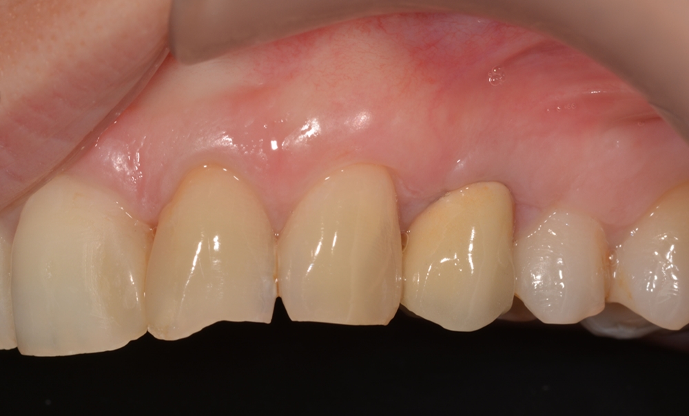
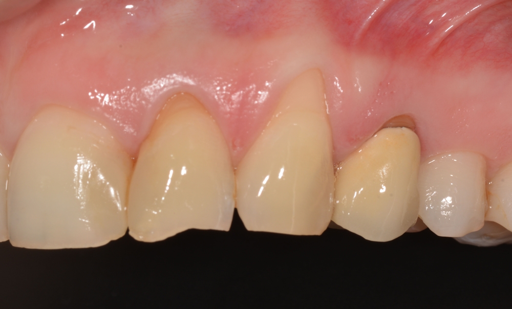
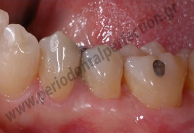
Diagnosis:Root recession in the first premolar upper right and non-carious cervical lesion that concerns both the root and the anatomical crown.
Treatment:Initially a cervical resin filling was fabricated in order to restore the root part of the cervical lesion (imitation of cementoenamel junction). Then a soft tissue graft was placed at the part of the cervical lesion corresponding to the anatomical crown root utilizing a variant of the original Zucchelli technique. The photo was taken 2 months after surgery and the root coverage is 100%.

CASE 12
Diagnosis: Υφίζηση στον κεντρικό τομέα κάτω αριστερά.
Treatment: Υποεπιθηλιακό μόσχευμα συνδετικού ιστού με τεχνική tunnel. Η τελική φωτογραφία λήφθηκε 6 μήνες μετά την χειρουργική και η κάλυψη της ρίζας είναι 100%.
CASE 11
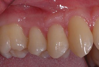
Diagnosis: Υφίζηση στον κυνόδοντα και τους προγομφίους άνω δεξιά και έλλειψη κερατινοποιημένων ιστών.
Treatment: Υποεπιθηλιακό μόσχευμα συνδετικού ιστού με μηλικά μετατοπιζόμενο κρημνό. Η φωτογραφία λήφθηκε δύο χρόνια μετά την χειρουργική και η κάλυψη είναι 100%. Παράλληλα επιτέυχθηκε και αύξηση του πάχους των κερατινοποιημένων ιστών
CASE 10
Diagnosis: Υφίζηση στον κεντρικό τομέα της κάτω γνάθου δεξιά. Η ασθενής έχει υποβληθεί σε ορθοδοντική.
Treatment: Υποεπιθηλιακό μόσχευμα συνδετικού ιστού με τεχνική tunnel. Η κάλυψη αγγίζει το 100% και το αποτέλεσμα είναι πολύ σταθερό μετά την πάροδο 5 ετών!!!
CASE 9
Diagnosis: Υφιζήσεις στους 4 τομείς της κάτω γνάθου με ταυτόχρονη έλλειψη κερατινοποιημένων ιστών.
Treatment: Ελεύθερο ουλικό μόσχευμα με σκοπό την αύξηση των κερατινοποιημένων ιστών καθώς η κάλυψη δεν θα ήταν εφικτή. Η φωτογραφία είναι 5 χρόνια μετά τη χειρουργική και η αύξηση των κερατινοποιημένων ιστών είναι θεαματική.
CASE 8
Diagnosis: Υφίζηση στον κεντρικό τομέα κάτω δεξιά σε νεαρή ενήλικη κοπέλα μετά από ορθοδοντική.
Treatment: Η τεχνική που επιλέχθηκε ήταν ελεύθερο ουλικό μόσχευμα για ταυτόχρονη κάλυψη της ρίζας αλλά και αύξηση των κερατινοποιημένων ιστών. Η λήψη της φωτογραφίας είναι 8 χρόνια μετά τη χειρουργική . Η κάλυψη τις ρίζας είναι 100% και η αύξηση των κερατινοποιημένων ιστών είναι θεαματική.
CASE 7
-
CASE 6
-
CASE 5
-
CASE 4
-
CASE 3
-
CASE 2
-
CASE 1
Recessions in upper premolar teeth and almost 100% root coverage after subepithelial connective tissue graft in a female smoker.